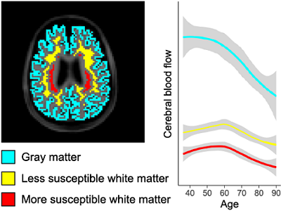
Focus 1: Cerebral hemodynamics in typical and abnormal aging
The human brain requires a constant supply of oxygen and other nutrients delivered through the bloodstream. Cerebral blood flow (CBF), which is a measure of perfusion, and arterial transit time (ATT), which is the time taken by arterial blood to reach the microvasculature, represent important markers of hemodynamic function. Our lab is interested in applying noninvasive MRI approaches to understand
(a) how aging impacts these measures in the brain,
(b) whether these effects differ in gray versus white matter, and
(c) the impact of various risk factors for cerebrovascular disease and dementia on hemodynamic measures.
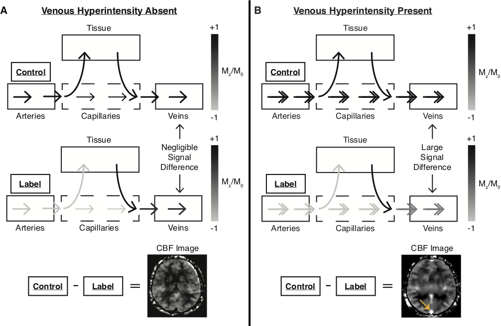
Focus 2:Noninvasive detection of altered capillary hemodynamics
Once oxygen-rich blood is delivered to the microvasculature, oxygen is offloaded from capillaries into brain tissue. The oxygen extraction fraction (OEF) represents the ratio of oxygen consumed by brain tissue to the oxygen delivered and is limited by microvascular flow patterns. Our lab is interested in developing noninvasive MRI approaches for characterizing
(a) the presence of such capillary flow disturbances that could lead to inefficient OEF,
(b) how inefficient OEF interacts with hypoperfusion mechanisms to incur hemodynamic impairment, and
(c) the degree to which these mechanisms contribute to white matter lesions in an aging population and individuals at risk for Alzheimer’s disease.
Meher R. Juttukonda, Manus J. Donahue, Spencer L. Waddle, Larry T. Davis, Chelsea A. Lee, Niral J. Patel, Sumit Pruthi, Adetola A. Kassim, and Lori C. Jordan.
Journal of Cerebral Blood Flow and Metabolism. 2021 Mar 41(3):546-560 | PMID: 32281458
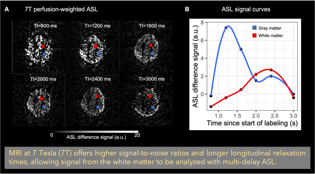
Focus 3:Imaging white matter perfusion with high-field MRI
White matter hemodynamic function has proven challenging to measure due in part to the lack of sensitive noninvasive methods for measuring associated physiology. Our lab is interested in developing MRI approaches at high-field (i.e., 7 Tesla) for characterizing the association between white matter lesion burden and white matter hemodynamic measures including
(a) perfusion,
(b) cerebrovascular reactivity, and
(c) oxygen extraction fraction.
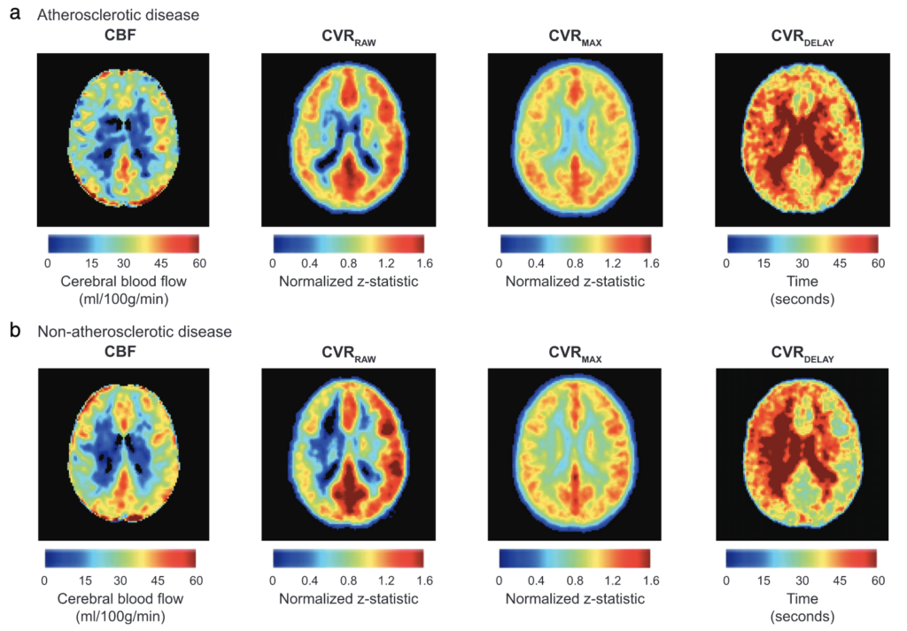
Focus 4:Hemo-metabolic function in cerebrovascular diseases
Cerebrovascular diseases often restrict oxygen availability to the brain through impairment to the hemo-metabolic equilibrium. Our lab is interested in applying noninvasive, clinically-relevant imaging approaches to understand how tissue-level physiology compensates for this impairment in patients with cerebrovascular diseases including
(a) sickle cell disease (SCD),
(b) arterial steno-occlusive disease, including atherosclerotic and moyamoya diseases,